Diagram Of Shoulder Bones / Glenohumeral Shoulder Joint Bones Movements Muscles Kenhub
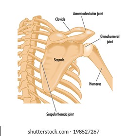
Diagram Of Shoulder Bones / Glenohumeral Shoulder Joint Bones Movements Muscles Kenhub. In human anatomy, the shoulder comprises the part of the body where the arm attaches to the torso. Diagram of the shoulder anatomy of shoulder ligament ideas anatomy lesson full hd wallpaper. Long bones are mostly located in the appendicular. Download a free preview or high quality adobe illustrator ai, eps, pdf and high resolution jpeg versions. Cheek bone (zygoma) upper jaw (maxilla). The main functions of shoulder bone are : It is made up of three bones: The shoulder muscles bridge the transitions from the torso into the head/neck area and.
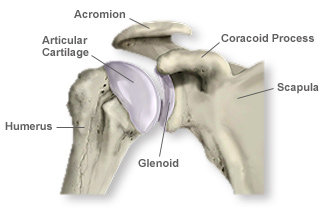
The transverse humeral ligament is not shown on this diagram. Joints hold your bones together and allow your rigid ball and socket joints, like your hip and shoulder joints, are the most mobile type of joint in the human body. The shoulder joint (glenohumeral joint) is a ball and socket joint between the scapula and the humerus. The human shoulder is made up of three bones:

(1) collar bone on the two sides of the next keep our shoulders apart.
Hand drawn realistic human bones. Cheek bone (zygoma) upper jaw (maxilla). There also are bands of fibrous connective tissue—the ligaments and the tendons—in intimate relationship with the parts of the skeleton. Bones » shoulder bones anatomy shoulder bone anatomy diagram human anatomy diagram categories: The shoulder bones, rib bones and hip bones ,are all joined to the backbone. When this type of cartilage starts. The bones of the shoulder consist of the humerus (the upper arm bone), the scapula (the shoulder blade), and the clavicle (the collar bone). It is made up of three bones: The main functions of shoulder bone are : The primary function of the shoulder girdle is to give strength and range of motion to the arm.
Labeled human shoulder bone anatomical vector illustration diagram poster. They allow you to swing your arms and. Ear wax normally comes out of your ear naturally so it's not a good idea to try and remove it yourself unless it is causing health problems (best to see your doctor first). The following diagram shows the structures related to shoulder joint. Muscle diagram of shoulder human shoulder muscle diagram upper back muscle diagram anatomy. Bones » shoulder bones anatomy shoulder bone anatomy diagram human anatomy diagram categories: Frozen shoulder occurs due to adhesive capsulitis, a disorder in which the capsule and the connective tissue surrounding the shoulder joint becomes inflamed and stiff, greatly restricting movement of shoulder.
The transverse humeral ligament is not shown on this diagram.
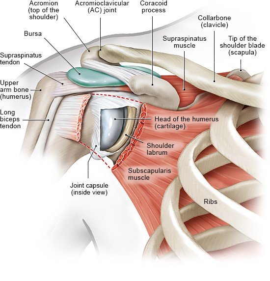
The shoulder joint is formed where the humerus (upper arm bone) fits into the scapula. The articulations between the bones of the shoulder make up the shoulder joints. Due to this, there is a hollow centre inside the backbone. Tutorials on the shoulder muscles (e.g rotator cuff muscles: This framework consists of many individual bones and cartilages. The following diagram shows the structures related to shoulder joint. Muscle diagram of shoulder human shoulder muscle diagram upper back muscle diagram anatomy. The shoulder is not a single joint, but a complex arrangement of bones, ligaments, muscles, and tendons that is better called the shoulder girdle. Shoulder joint is the most mobile joint of the human body. The shoulder muscles bridge the transitions from the torso into the head/neck area and.
The bones of the shoulder consist of the humerus (the upper arm bone), the scapula (the shoulder blade), and the clavicle (the collar bone). Shoulder diagram illustrations & vectors. The first type is the white cartilage on the ends of the bones (called articular cartilage) which allows the bones to glide and move on each other. Other important bones in the shoulder include shoulder joint of human body anatomy infographic diagram with all parts including bones ligaments muscles bursa cavity capsule cartilage membrane for medical science education and health care. (1) collar bone on the two sides of the next keep our shoulders apart. Scapula (= 'shoulder blade' or 'shoulder bone') is a bone of the human body. It is made up of three bones: We discuss their function, the different types of bones in the human body, and the cells that are involved. Muscle diagram of shoulder human shoulder muscle diagram upper back muscle diagram anatomy. Neck vertebrae (7) (cervical vertebrae).
Very soon we'll move on to muscles!
All of your bones, except for one (the hyoid bone in your neck), form a joint with another bone. There are two kinds of cartilage in the joint. Very soon we'll move on to muscles! 9 photos of the shoulder bones anatomy diagram. Long bones are mostly located in the appendicular. The articulations between the bones of the shoulder make up the shoulder joints. The largest bone in the human body is the thighbone or femur, and the smallest is the stapes in the middle ear, which are just 3 millimeters (mm) long. These are the supraspinatus, infraspinatus, teres. The main functions of shoulder bone are : Simple structure of the clavicle. Lower jaw (mandible) collar bone. It is made up of three bones: Most relevant best selling latest uploads.
All of your bones, except for one (the hyoid bone in your neck), form a joint with another bone diagram of shoulder. Shoulder diagram to mainly explain you about how your shoulder work and to describe every inner part of your shoulder including muscles, joints, and every human has a body part called shoulder.

Click now and learn everything about its anatomy and function at kenhub!

These are the supraspinatus, infraspinatus, teres.

Download 708 shoulder diagram stock illustrations, vectors & clipart for free or amazingly low rates!

Very soon we'll move on to muscles!
 fits into the scapula.")
Each vertebra has a hole in it.

Scapula (= 'shoulder blade' or 'shoulder bone') is a bone of the human body.

The shoulder joint (glenohumeral joint) is a ball and socket joint between the scapula and the humerus.

The rotator cuff muscles are four muscles that form a musculotendinous unit around the shoulder joint.

These are the supraspinatus, infraspinatus, teres.

In human anatomy, the shoulder comprises the part of the body where the arm attaches to the torso.

Consisting of the clavicle (collar bone) and scapula (shoulder blade), the pectoral girdle forms the attachment point between the arm and the chest.

Shoulder problems including pain, are one of the.

Each vertebra has a hole in it.
, the scapula (the shoulder blade), and the clavicle (the collar bone).")
It is made up of three bones:

Long bones are mostly located in the appendicular.

Bones » shoulder bones anatomy shoulder bone anatomy diagram human anatomy diagram categories:

Long bones are mostly located in the appendicular.

These are the supraspinatus, infraspinatus, teres.

Simple structure of the clavicle.

Very soon we'll move on to muscles!

Other important bones in the shoulder include shoulder joint of human body anatomy infographic diagram with all parts including bones ligaments muscles bursa cavity capsule cartilage membrane for medical science education and health care.

We discuss their function, the different types of bones in the human body, and the cells that are involved.

9 photos of the shoulder bones anatomy diagram.
The first type is the white cartilage on the ends of the bones (called articular cartilage) which allows the bones to glide and move on each other.

9 photos of the shoulder bones anatomy diagram.
 is a ball and socket joint between the scapula and the humerus.")
Cheek bone (zygoma) upper jaw (maxilla).

The main functions of shoulder bone are :

Diagram of the shoulder anatomy of shoulder ligament ideas anatomy lesson full hd wallpaper.
 long.")
The scapula is a large, flat triangular bone with three processes called the acromion, spine and coracoid process.


Posting Komentar
0 Komentar